
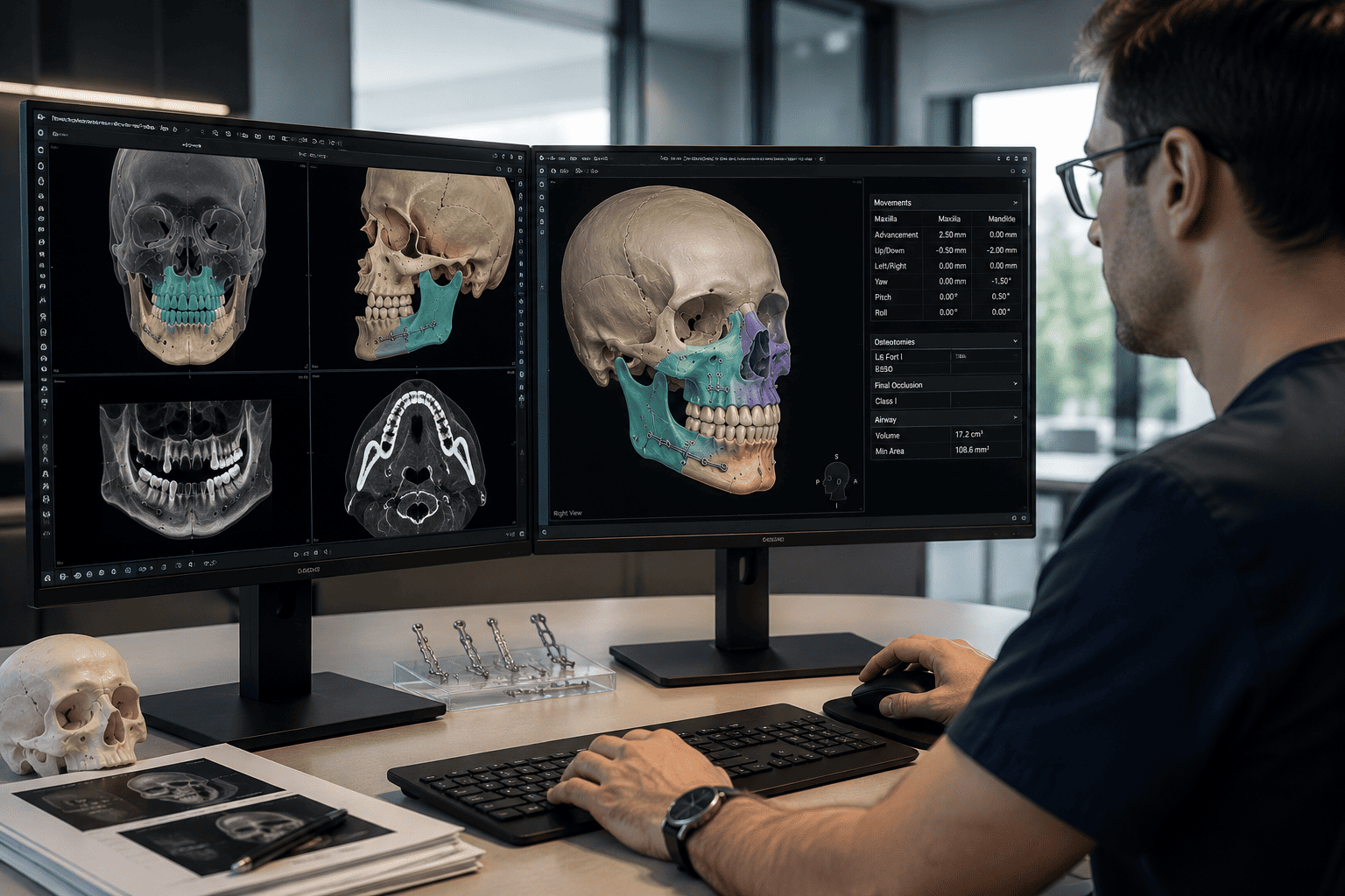
In maxillofacial surgery, precise planning is not simply about deciding how far a bone should be moved.
It requires understanding a three-dimensional anatomy, defining osteotomies, anticipating how the bone segments will relate to one another once mobilized, assessing occlusion, recognizing anatomical structures that must be preserved and, ultimately, ensuring that what has been planned can be reproduced during the procedure.
3D virtual planning has profoundly transformed this process.
Starting from imaging studies and digital records, it is possible to reconstruct the patient's craniofacial anatomy, study the deformity in three dimensions, simulate different surgical strategies and manufacture devices designed to transfer certain components of the plan to the operating room.
But reducing this technology to the ability to "see the surgery before performing it" would oversimplify its true scope.
Its value lies in something more important: turning complex anatomical information into quantifiable surgical decisions and enabling a controlled transfer of those decisions to the operative field.
What is 3D virtual planning in maxillofacial surgery?
Virtual surgical planning—often called Virtual Surgical Planning or VSP—is a digital workflow through which a three-dimensional representation of the patient's anatomy is built and used to analyze, simulate and transfer a surgical procedure.
It is not simply a three-dimensional reconstruction of a CT scan.
A reconstruction allows visualization.
Planning requires interpreting, deciding and transferring.
The process can integrate:
- computed tomography or cone-beam computed tomography;
- DICOM files from the radiological study;
- intraoral scanning or digitization of dental models;
- clinical photographs;
- three-dimensional models;
- segmentation of anatomical structures;
- simulation of osteotomies;
- virtual movements of bone segments;
- occlusion analysis;
- CAD/CAM surgical splints;
- cutting or positioning guides;
- 3D-printed anatomical models;
- and, in certain procedures, patient-specific plates or implants.
The result is a virtual model on which an operation can be studied before it is physically performed.
However, 3D planning does not turn a complex procedure into an automatic operation. The software represents the anatomy and executes the instructions of the plan; the indication, the objectives and the decisions remain clinical.
The first step is not the software: it is the diagnosis
The quality of a virtual plan depends on the quality of the clinical question it is trying to answer.
Before segmenting structures or simulating movements, a diagnosis must exist.
In orthognathic surgery, for example, it is necessary to determine where the deformity is located, which component is maxillary, mandibular, dentoalveolar, transverse, vertical or asymmetric, and what functional and facial goals are being pursued.
In facial trauma, one must understand the alteration of the bony architecture caused by the fracture.
In reconstructive surgery, it must be established what structure is missing, what must be preserved and what geometry needs to be reconstructed.
Technology can provide an extraordinarily detailed representation of the anatomy, but more information does not automatically translate into a better diagnosis.
Virtual planning gains true value when that information modifies or refines a surgical decision.
From image acquisition to the virtual patient
The process begins with obtaining sufficiently precise anatomical information.
Three-dimensional radiological imaging
CT scanning provides volumetric information about the craniofacial skeleton.
Data is usually stored in DICOM format and allows the structures to be reconstructed in three dimensions.
In orthognathic surgery, CBCT is widely used for this purpose. Reviews on the accuracy of virtual planning describe precisely the combination of CBCT with digital dental records as one of the most widely used protocols.
The quality of this initial acquisition matters.
Movement during the scan, metal artifacts, dental restorations, orthodontic appliances or inadequate resolution can affect certain regions of the reconstruction.
A visually convincing three-dimensional model does not by itself guarantee that all of its data have the same accuracy.
Digital dental records
Dentition represents a particular challenge.
Dental surfaces obtained directly from a CT scan may show artifacts or insufficient definition for certain occlusal objectives.
For this reason, intraoral scans or digitization of models can be used to obtain more precise dental surfaces.
These data are subsequently registered with the radiological information.
The result is a composite model: the bony anatomy comes mainly from the volumetric image, while the high-resolution dental anatomy may come from optical scanning.
The accuracy of the registration between both data sets is essential, because an error at this stage can propagate to later steps.
Photography and soft tissues
Maxillofacial surgery does not deal solely with bones.
In orthognathic surgery, modifying the position of the skeleton also modifies the lips, cheeks, chin, nasal base and other facial structures.
Clinical photographs and, when available, other three-dimensional surface records can complement the analysis.
Current software allows soft-tissue predictions to be made, but these must be interpreted correctly: they are computational estimates, not a guaranteed representation of the final facial outcome.
Soft tissues respond biologically, and not entirely linearly, to skeletal movements.
Segmentation: turning an image into operable anatomy
A CT scan contains continuous volumetric information. To work surgically with it, the relevant structures must be identified and separated.
This process is called segmentation.
Depending on the procedure, it can allow the following to be individualized:
- maxilla;
- mandible;
- dentoalveolar segments;
- chin;
- orbits;
- zygomatic complex;
- reconstructed structures;
- bone defects;
- and other anatomical regions of interest.
From this segmentation, manipulable three-dimensional models are generated.
This step is fundamental because it constitutes the transition between a diagnostic image and a model on which a procedure can be simulated.
It is also a possible source of error.
Automated algorithms have considerably accelerated the process, but certain anatomical areas, artifacts, thin bony margins or pathological situations may require review and correction.
Automation reduces workload. It does not eliminate the need to validate the anatomy that will be used for planning.
Virtual surgery: simulating before intervening
Once the model has been built and validated, the osteotomies can be simulated.
In orthognathic surgery, for example, a Le Fort I osteotomy, a sagittal split mandibular osteotomy, a genioplasty or combinations of procedures can be virtually reproduced.
The resulting segments can then be moved along the three spatial axes and rotated.
This allows the analysis of movements that are more difficult to understand simultaneously in an exclusively two-dimensional plan:
- advancement and setback;
- impaction and downward movement;
- lateral translation;
- yaw corrections;
- pitch corrections;
- roll corrections;
- rotations of the maxillomandibular complex;
- asymmetric corrections.
The difference is conceptually important.
A facial deformity is three-dimensional. An asymmetry, for example, can rarely be adequately explained by a single linear distance.
3D planning allows the study of how position, orientation and rotation interact simultaneously.
It is not about moving bones until they "look right"
A rigorous surgical simulation should not become an exercise in visual manipulation.
Every movement must respond to an objective.
In orthognathic surgery, the final position of the jaws must integrate, among other elements:
- occlusal relationship;
- midlines;
- dental exposure;
- occlusal plane;
- symmetry;
- facial proportions;
- anteroposterior position;
- vertical dimension;
- transverse relationships;
- temporomandibular joint;
- available anatomy;
- expected stability;
- and the anticipated response of the soft tissues.
One movement can improve one variable and worsen another.
This is why the ability to simulate multiple scenarios is useful: it allows the geometric consequences of different strategies to be studied before selecting one.
Virtual planning is not about finding the most spectacular movement, but the movement most consistent with the diagnosis.
Critical anatomy: planning also means knowing where not to intervene
One of the conceptual advantages of the three-dimensional environment is the ability to analyze the relationship between osteotomies and certain anatomical structures.
Depending on the procedure, the mandibular canal, dental roots, orbital walls, paranasal sinuses, vascular structures or other anatomical elements may be relevant.
Planning can help study:
- osteotomy trajectories;
- bone availability;
- interferences between segments;
- planned screw position;
- the relationship between fixation devices and nearby structures;
- the geometry of a reconstructive defect.
This is especially important in anatomies altered by severe deformities, trauma, previous surgeries or resections.
Planning is not only about determining where a segment will end up.
It is also about determining how it can get there in an anatomically reasonable way.
From virtual to physical: the real problem of transfer
There is a fundamental distinction between planning with precision and operating with precision.
A virtual plan can be geometrically excellent and yet lose accuracy during its transfer to the patient.
For this reason, one of the central issues in computer-assisted surgery is the mechanism by which digital coordinates are physically reproduced during the procedure.
This transfer can be carried out using different strategies.
CAD/CAM surgical splints
In orthognathic surgery, once the relationship between the jaws has been determined virtually, digital splints can be designed and manufactured using CAD/CAM technologies.
Splints allow certain occlusal relationships and planned positions to be transferred to the operating room.
They represent an advance over traditional model surgery processes, but they still depend on intraoperative references and have inherent limitations, particularly in controlling certain spatial dimensions.
Cutting and positioning guides
In selected procedures, specific devices can be designed that adapt to the patient's anatomy.
These guides can help reproduce:
- osteotomy locations;
- drill holes;
- reference positions;
- orientation of certain segments.
Their usefulness depends on the guide fitting stably to the actual anatomy, and on all previous steps—imaging, segmentation, design and manufacturing—having been carried out correctly.
Patient-specific plates and devices
In certain cases, osteosynthesis systems specific to the anatomy and the planned movement can be designed.
The concept differs from adapting a standard plate during the operation.
The desired final position is incorporated into the design of the fixation system and can be combined with cutting or drilling guides.
Clinical studies and randomized trials have investigated whether these specific systems can improve transfer accuracy compared with CAD/CAM splints. The results are promising, although their use should be evaluated according to the procedure, complexity, cost and available evidence.
Surgical navigation
Another strategy involves using navigation systems that relate the patient's actual anatomy to the virtual model during the procedure.
This allows the position of certain structures or instruments to be checked intraoperatively.
Navigation has particularly interesting applications in certain craniofacial regions, although it requires equipment, precise registration and a specific workflow.
What does virtual planning really add compared with conventional planning?
For decades, complex maxillofacial surgeries were performed using conventional planning methods, including cephalometric analysis, plaster models, articulators and model surgery.
The arrival of virtual planning does not retroactively make those methods incorrect.
What changes is the amount and nature of the information available.
Three-dimensional planning allows:
- simultaneous study of the three spatial planes;
- visualization of complex asymmetries;
- quantification of translations and rotations;
- simulation of different strategies;
- analysis of certain anatomical interferences;
- integration of digital records;
- design of personalized devices;
- subsequent comparison of the achieved result with the original plan.
The available systematic reviews find that virtual planning can achieve high accuracy and reproducibility. One review on orthognathic surgery found mean translational discrepancies of less than 2 mm between planning and outcome for the maxilla and mandible across the studies analyzed.
However, the comparative literature calls for caution.
Reviews of clinical trials have found heterogeneous results on some accuracy measures compared with conventional planning, although there are advantages in certain parameters and a trend toward better correspondence between plan and outcome.
It would therefore be incorrect to claim that "3D means absolute accuracy."
The more clinically rigorous statement is different: virtual planning provides considerably more powerful tools for analyzing, simulating, quantifying and transferring a three-dimensional surgery.
Where is it especially useful?
Virtual planning can be applied to different areas of oral and maxillofacial surgery.
Orthognathic surgery
This is probably one of its most developed applications.
It allows dentofacial deformities to be studied three-dimensionally, maxillomandibular movements to be simulated, asymmetries to be analyzed, and splints or personalized systems to be manufactured to transfer the plan to the operating room.
Craniofacial asymmetries
Asymmetric deformities are particularly well suited to three-dimensional analysis because they involve simultaneous differences between both sides of the face.
The ability to compare structures, quantify deviations and analyze rotations provides information that is difficult to condense in two-dimensional studies.
Facial trauma
In certain complex fractures or traumatic sequelae, the residual anatomy, the contralateral side or reconstructed models can be used to study the restoration of facial architecture.
Maxillofacial reconstruction
After tumor resections or complex bone loss, virtual planning can help define the defect, design the reconstruction and plan the geometry of the bone segments.
In microvascular reconstructions, for example, guides can be designed to facilitate certain osteotomies of the graft and its adaptation to the defect.
Implant surgery and personalized reconstructions
In certain scenarios, digital models allow the design of implants or components specific to a particular anatomy.
However, the indication and the complexity of the digital workflow always depend on the clinical problem being addressed.
Does 3D planning improve accuracy?
The correct answer requires nuance.
The available literature shows that virtual planning can be clinically reproduced with a high degree of accuracy.
A systematic review on orthognathic surgery found mean discrepancies of less than 2 mm in maxillary and mandibular translations across the included studies, although there was methodological variability.
Meta-analyses and subsequent reviews have continued to show favorable results for virtual planning across different parameters, but have also pointed out heterogeneity between studies and the absence of a universal method for measuring accuracy.
Furthermore, final accuracy does not depend on the software alone.
There is a chain of accuracy:
image acquisition → segmentation → registration → planning → design → manufacturing → intraoperative adaptation → surgical execution.
An error introduced at any of these steps can be carried forward to the next.
For this reason, discussing digital accuracy without analyzing the complete workflow can be misleading.
Can the facial outcome be predicted exactly?
No.
Software can simulate the displacement of soft tissue associated with skeletal movements, and these tools are useful for planning and communication.
But a virtual prediction should not be confused with a promise of a result.
Soft-tissue response depends on numerous factors:
- thickness and individual characteristics of the tissues;
- magnitude and direction of the bone movement;
- muscle tone;
- age;
- healing;
- edema;
- associated procedures;
- biological adaptation.
Furthermore, different facial regions do not respond identically to movement of the underlying skeleton.
Simulation should be used as an analytical tool, not as an aesthetic guarantee.
Does virtual planning reduce surgical time?
Some comparative studies and reviews have found reductions in intraoperative time with virtual workflows.
However, evaluating only operating-room time gives an incomplete picture.
Some of the work that used to be carried out through laboratory procedures or during the operation can be shifted to preoperative digital stages.
True efficiency must consider the entire process:
- acquisition;
- processing;
- planning;
- planning meetings;
- design;
- manufacturing;
- surgery.
Furthermore, the more personalized the solution—guides, specific plates, implants—the greater the required prior preparation may be.
The fundamental advantage should not be measured solely in minutes saved, but in the ability to anticipate and control the procedure.
What are the limitations of 3D virtual planning?
A mature surgical technology must also be analyzed in terms of its limitations.
Dependence on data quality
A deficient image or an incorrect registration compromises the model.
Segmentation errors
Anatomical boundaries can be misinterpreted, especially in the presence of artifacts or very thin structures.
Registration errors
The integration between CT scan, dental scanning and other records must be precise.
Limitations of soft-tissue prediction
Mathematical models do not fully reproduce biological variability.
Imperfect transfer to the operating room
The existence of a precise plan does not guarantee that it can be reproduced exactly.
Cost and availability
Software, engineering, manufacturing of specific devices and equipment can increase costs compared with certain conventional workflows.
Comparative reviews have specifically identified higher financial cost as one of the current disadvantages of some virtual planning protocols.
Learning curve
The ability to manipulate digital models is not the same as understanding three-dimensional surgery.
There is a learning curve, both technological and clinical.
What risks does the technology add?
Virtual planning does not replace or eliminate the risks inherent to the surgical procedure being performed.
In addition, it introduces possible sources of error specific to the digital workflow:
- incorrect data acquisition;
- defective segmentation;
- imprecise fusion of records;
- design errors;
- manufacturing errors;
- inadequate fit of a guide;
- use of incorrect anatomical references;
- discrepancies between the planned anatomy and the anatomy found during surgery.
For this reason, personalized devices must be verified, and the surgeon must retain the ability to modify the plan when the intraoperative situation requires it.
A virtual plan should not become a surgical obligation if the actual anatomy shows that something different must be done.
What happens after surgery?
The digital workflow does not necessarily end once the procedure is finished.
When clinically indicated, postoperative images can be compared with the virtual plan.
Through three-dimensional superimposition, differences can be analyzed between:
- planned position;
- achieved position;
- translations;
- rotations;
- symmetry;
- position of segments or reconstructions.
This capability has clinical importance, but also academic importance.
It allows the accuracy of a technique to be objectively evaluated, the origin of deviations to be studied, and planning and transfer protocols to be progressively improved.
Digital surgery, in this sense, does not only allow planning.
It also allows measurement.
Technology and surgical judgment: which should guide which
Technological sophistication can create a false sense of certainty.
A three-dimensional reconstruction can be extremely visually convincing. A movement can be expressed in tenths of a millimeter. A plate can be manufactured specifically for a patient.
None of this guarantees that the initial decision was correct.
There can be a technically perfect plan for a conceptually mistaken strategy.
This is why the appropriate sequence is:
diagnosis → objectives → strategy → simulation → validation → transfer → execution → evaluation.
Not:
software → simulation → surgery.
This difference is essential.
3D virtual planning reaches its maximum value when it amplifies the surgeon's analytical capacity, not when it attempts to replace it.
Frequently asked questions about 3D virtual planning
Do all maxillofacial surgeries need 3D planning?
No. Its usefulness depends on the procedure and the complexity of the case. There are procedures in which a conventional workflow may be sufficient, and others in which three-dimensional analysis provides decisive information.
Does 3D planning mean the surgery will be exactly the same as the simulation?
No. The simulation establishes an objective and a strategy. The anatomy, the transfer technique and the intraoperative circumstances determine the final correspondence between planning and outcome.
Can you know exactly what the face will look like before orthognathic surgery?
No. An approximate prediction of soft-tissue changes can be made, but an exact reproduction of the facial outcome cannot be guaranteed.
What is the difference between a CAD/CAM splint and a personalized plate?
A surgical splint helps transfer certain planned relationships, especially occlusal ones. A patient-specific plate can be designed to fit a previously planned anatomy and bone position. These are different transfer strategies and are not interchangeable in every case.
Are 3D printing and virtual planning the same thing?
No. Virtual planning is the process of digital analysis and simulation. 3D printing is a manufacturing technology that can be used to produce models, guides or other devices derived from that planning.
Does virtual planning eliminate human error?
No. It changes where certain errors can occur and allows certain variables to be controlled with greater precision, but it still depends on data acquisition, processing, interpretation and surgical execution.
Can the plan be modified during the operation?
Yes. Planning establishes a preoperative strategy, but the surgeon must be able to adapt it if intraoperative findings require it.
Is it more accurate than conventional planning?
The available evidence shows high accuracy and advantages in different parameters, but the comparisons are not uniform across all studies. The magnitude of the advantage also depends on the procedure and the system used to transfer the plan.
From the image to the operating room
The main contribution of 3D virtual planning is not producing more sophisticated images.
It consists of creating continuity between diagnosis, strategy and execution.
The anatomy obtained through medical imaging becomes a model. The model allows the deformity to be studied. Osteotomies and movements can be simulated on it. These movements can be turned into splints, guides, fixation systems or navigation references. Finally, the result can be compared with the originally planned objective.
This path—from image to operating room and from the operating room back to analysis—represents one of the most significant changes in contemporary maxillofacial surgery.
But technology does not change one fundamental principle.
Accuracy begins long before a guide or plate is manufactured. It begins by correctly defining what must be done and why.
Virtual planning can make surgery more measurable, reproducible and controllable.
Clinical judgment continues to determine which surgery is worth planning.
Selected scientific references
- Alkhayer A, Piffkó J, Lippold C, Segatto E. Accuracy of virtual planning in orthognathic surgery: a systematic review. Head & Face Medicine. 2020;16:34.
- Starch-Jensen T, Hernández-Alfaro F, Kesmez Ö. Accuracy of Orthognathic Surgical Planning using Three-dimensional Virtual Techniques compared with Conventional Two-dimensional Techniques: a Systematic Review. Journal of Oral & Maxillofacial Research. 2023;14(1):e1.
- Strujak G, Marlière DAA, de Lima Medeiros Y, et al. Virtual Versus Conventional Planning in Orthognathic Surgery: A Systematic Review and Meta-analysis. Journal of Maxillofacial and Oral Surgery. 2024;23(2):219–228.
- Saigo L, Schrader F, Rana M, Wilkat M. 3-Dimensional accuracy of navigation-guided bimaxillary orthognathic surgery: A systematic review and meta-analysis. Journal of Cranio-Maxillofacial Surgery. 2024;52(11):1274–1287.
- Gupta S, et al. Effectiveness of Traditional and Virtual Surgical Planning in Orthognathic Surgery: A Systematic Review and Meta-Analysis. 2025.